How to See like Bats
Sound is the vocabulary of nature.
Pierre Schaeffer
You are doing your daily ward round, and the nurse calls you saying the patient who underwent hernia repair a few hours earlier has lower abdominal pain. What would you do?
Or, maybe, you are on call and a hemodynamically unstable patient after a blunt abdominal trauma arrives. What could you do to help you decide the best management?
And, what about the patient you operated on for acute cholecystitis a couple of days back, who has fever and upper right quadrant abdominal pain?
There are many clinical decisions that can be solved with just a simple and fast exam, and this exam can be performed at the bedside… Ultrasound!!!
Even if we are surgeons and not radiologists, ultrasound is a powerful tool that can help us clarify and speed up difficult and/or doubtful decisions…
Do you want to see your patient in depth, but you don’t know how to do it? Let’s see it together!
Basics of US
If you have already listened about US but you don’t know anything about its physical and technical principles, here you are an explanation for dummies of how it works.
1. Production and detection of ultrasound waves
Ultrasound is a wave that travels through an elastic medium with a frequency range of 1-20Mhz.
Sound waves are characterized by intensity, wavelength, frequency, and velocity.
Diagnostic ultrasound is based on the pulse-echo principle. Piezoelectric crystals (the smallest functional unit of a transducer) have a double function: convert electrical oscillations into vibration,s generating a pulse of sound, and listen to the echoes reflected from interfaces.
2. Propagation of sound
When a US wave encounters an interface, part of the sound bounces back with the same angle (reflection), part of it is converted into heat (absorption), part of it is scattered away from the object in random directions (scatter) and part of it continues to go through the second medium (refraction), as shown in the figure below.
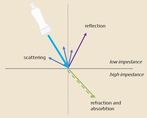
Impedance is the resistance of the medium to the propagation of sound; the higher it is between two media, the greater is the portion of the sound that will be reflected.
In real life:
- Air: reflects ultrasound almost completely, obscuring the visualization of underlying structures.
- Bone: absorbs and reflects much of the ultrasound energy, creating an acoustic shadow behind it.
- Liquid: do not reflect ultrasound and will appear black (anechoic) with a posterior reinforcement.
3. Production of an image
The returning sound brings back two principal information:
- The time it takes to come back, indicating the location of the reflecting interface
- The intensity of the echoe, demonstrating the hardness of the media
Ultrasound offers a large variety of different imaging modes:
- A-mode: the earliest US mode showed returning echoes in a one dimensional, graphical format.
- B-mode: “B” or Brightness mode represents the amplitude peaks seen in A-mode as dots or pixels of varying brightness. US systems can send sequential ultrasound pulses out in different directions to form multiple image lines. This process is completed quickly and repeatedly, creating the typical ultrasound images.
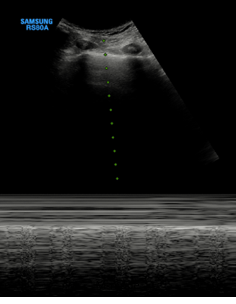
- M-mode: displays the echo data for a single image line over time. It is used to display moving structures.
4. Artifacts
Artifacts are “fake images” that do not correspond to anatomical structures, but are the result of US propagation in tissues.
A. Noise: it is an artifact from diffuse reverberation from tissues placed before a liquid-filled formation (e.g. bladder), and it can be reduced by lowering the gain.
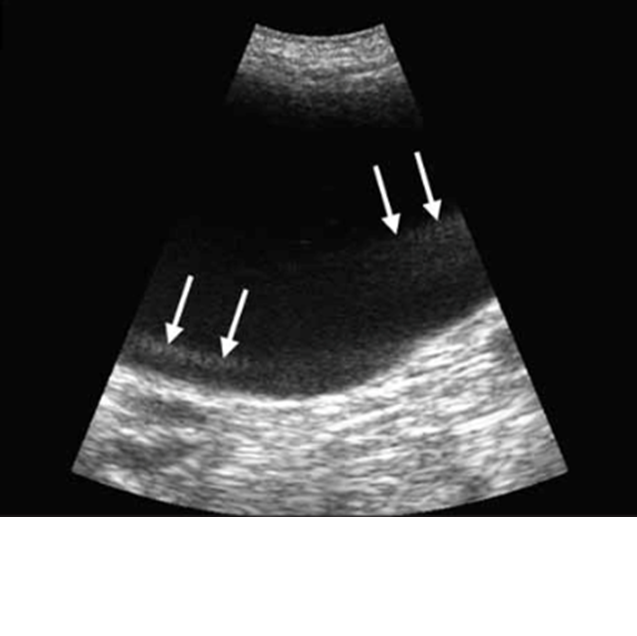
B. Shadow: it occurs when ultrasound encounters a highly attenuated structure (for example, a stone). This is the case with air or gas, bones, stones, and calcifications. The structures behind the stone appear dark or absent.
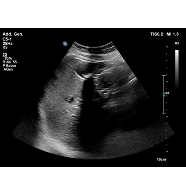
C. Posterior enhancement: this is characteristic of fluid-filled structures. Fluid attenuates sound much less than the surrounding tissue, so when ultrasound waves reach the opposite wall, where they are reflected, they maintain the same intensity as they had at the entrance and therefore provide a more intensely echogenic image of the surrounding areas, which are at the same depth but are hit by lower intensity ultrasound waves because they have already undergone dispersion upstream.
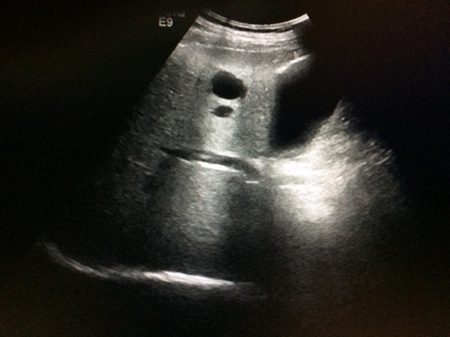
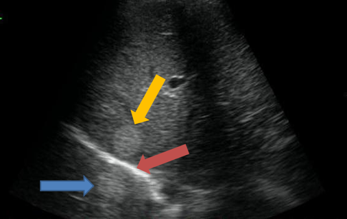
D. Mirror-image: it is seen when there is a highly reflective surface (e.g. diaphragm) in the path of the primary beam. A virtual image of the real object forms behind the highly reflective interface.
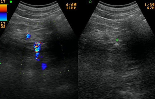
E. Comet-tail: it is typically seen in a situation when a small, highly reflective (usually calcific renal calculi) object is interrogated. With color Doppler, a twinkling artifact occurs, and immediately deep to the object, a tail of a linear, aliased color band extends away from the probe.
Well, that’s cool… but how to?!
Now that you know all the basics, let’s go through how to do!
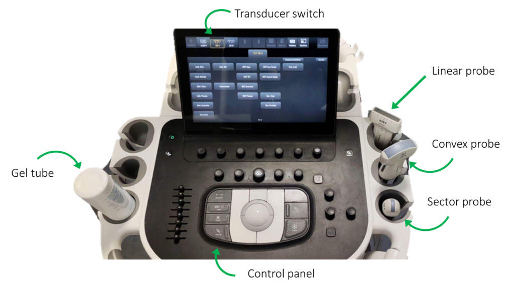
First, the equipment:
- Control panel
- Probe
- Monitor
Gel is fundamental because it cancels the resistance that air offers to the propagation of ultrasound through the skin.
Control panel
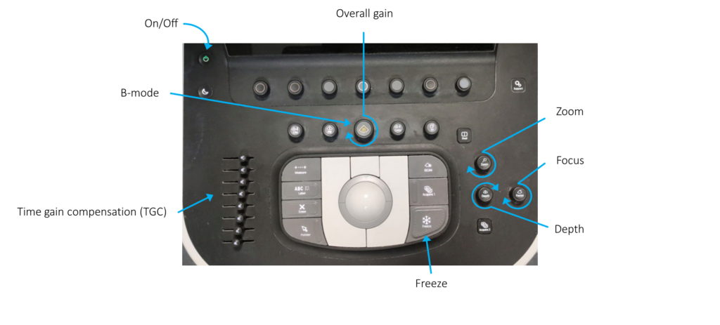
- Overall gain: amplifies echoes over the entire depth, affecting image brightness
- Zoom: enlarges an image section
- Focus: concentrates the beam of ultrasound on a target deep
- Depth: narrows or widens the image field
- Freeze: freeze the scan and allow to save an image
- TGC: amplifies echoes according to their depth
Probes
1. Types
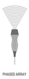 |  | 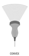 | |
| Beam shape | Fan shaped | Rectangular | Fan shaped |
| Frequency | Low | High | Low |
| Penetration | Deep | Superficial | Deep |
| Resolution | Low | High | Low |
| Best for | Small acoustic window | Superficial structures | Deep and wide fields |
2. Scans


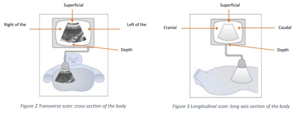
Every probe has an orientation marker on one side. Conventionally, the marker is on the LEFT side of the screen, indicating the right of the patient in transverse scans and the top in longitudinal scans.
In cardiac mode, it is on the RIGHT side of the screen.
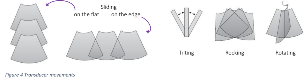
3. Transducer movements
We hope you found this post intriguing and useful. We really believe US is able to change and improve the way you manage your patients.
See you next time…
References
- Zago M. Essential US for Trauma: E-FAST. 1st Ed. Milan: Springer-Verlag; 2014
- Hofer M. Ecografia – Manuale didattico per l’esecuzione e l’interpretazione delle immagini. 10th Ed. Roma: Verduci Editore; 2021
How to Cite This Post
Germiniasi G, Cioffi SPB, Marrano E, Bellio G. How to See Like Bats. Surgical Pizza. Published on February 8, 2026. Accessed on October 18, 2026. Available at [https://surgicalpizza.org/imaging/how-to-see-like-bats/].