Newton and the Palm Tree: The Awful Coconut Law
It’s all in your head, Alice.
Lewis Carroll
Welcome back guys…
Let’s not lose time, and go straight to our daily (or monthly) clinical case…
You’re on call while all your friends are enjoying a sunny early summer day at the beach (we know we are cruel but that’s reality…) and a message on your beeper informs you a polytrauma patient is coming. At the bay (the one in the hospital, not that at the seaside), the nurse tells you what is coming:
A – 16 yo boy
T – 30 mins ago, arriving in 10 mins
M – Fall with a kickscooter in a skate park without a helmet
I – Mild headache
S – BP 145/90mmHg, Sat 98% in room air, HR 85 bpm R
T – Cervical collar, spinal table
You have a whole skilled team ready to help: two first-year residents of anesthesiology and general surgery, and a third-year resident of urology. Hell yes, it’s your lucky day! And the nursing team is made of 3 senior trauma nurses, they have your back!
Right after informing the team about the resuscitative plan the patient arrives: easy peasy, like he’s tanning on a beach…
A – Open and stable, central trachea, cervical collar on;
B – Normal bilateral breath sounds, nothing to report, Sat 98% in room air;
C – BP 140/85mmHg, HR 80 bpm, the abdomen is soft, the pelvis is stable, legs and arms are fine, no external bleeding;
D – GCS 13 (E3V4M6), no lateralizing signs, pupils are unequal but reactive to light equally.
Introduction, Anatomy & Physiology
Traumatic brain injuries (TBI) are extremely frequent and when we speak about polytrauma patients they are almost inevitable… The patient hits the head against something. Surely, we need to say that almost 80% of the TBI are of mild severity and with few consequences.
But what is so special about TBI?
Anatomically speaking the head is a closed and unexpandable space thanks to the skull, which protects the brain but doesn’t allow any adjustment of size. In the head, we find the scalp, skull, meninges (dura, arachnoid, and pia mater), brain, blood vessels (arterial and venous), and ventricular system with cerebrospinal fluid (CSF).
The CSF fills the ventricles and the meningeal’s spaces (subdural and subarachnoid space).
Clearly, this whole system needs to auto-regulate itself in a very complicated way due to the hundreds of parameters in play (e.g. arterial pressure, intracranial pressure, intracranial flow, pCO2, glucose, pO2….). There are several reasons why the intracranial pressure may rise, but here we are speaking about TBI so we mainly have 2 reasons: mass effect due to bleeding, or diffuse brain swelling because of diffuse axonal damage. Both of them will lead to an increased intracranial pressure (ICP) that the head will have to fight against.
Thanks to the Monro-Kellie doctrine, we know that the mass effect created by bleeding is compensated by a rapid outflow of CSF and venous blood. This will make the intracranial pressure stable for a while. However, when the mass is occupying more space, the system will rapidly decompensate! That is why an epidural hematoma may let the patient awake until the end (“they talk and die!” but we will get there).
The intracranial pressure (ICP) is normal at around 10 mmHg, it is worrying (abnormal) above 20 mmHg and it is extremely dangerous over 40 mmHg!
When we speak about cerebral perfusion pressure (CPP) we just need to remember that is the difference between the mean arterial pressure (MAP) and the ICP:
CPP = MAP – ICP
Fortunately, when the MAP is between 50 and 150 mmHg, thanks to vasoconstriction and vasodilation, our brain system can autoregulate itself to maintain the CPP stable.
Obviously, if the MAP is too low the brain will suffer ischemia, and if it’s too high there will be diffuse swelling.
The pCO2 and the pO2 will also have a part in regulating the vasoconstriction or dilation and we may use those to our advantage in some specific conditions (we’ll see them shortly).
Classification
How do we have to classify the TBI?!… We can classify them according to their morphology and severity. Unfortunately, giving just one classification may not be a complete description of the TBI.
The morphological classification is:
- Skull fractures
- Vault
- Linear vs. stellate
- Depressed/non-depressed
- Basilar
- With/without CSF leak
- With/without seventh nerve palsy
- Vault
- Intracranial lesions
- Focal
- Epidural
- Subdural/Subarachnoid
- Intracerebral
- Diffuse
- Concussion
- Multiple contusions
- Hypoxic/ischemic injury
- Axonal injury
- Focal
Let us say we will not cover all the lesions, but only the intracranial ones with a few words… Obviously, when we evaluate a trauma patient, the information we need to describe the TBI from the morphology point of view will come from the secondary survey and the TC images/report. So, we are speaking about a “stable” patient (at least from the ABC point of view, D may be still pending).
Focal lesions are hematomas that develop in between the meninges, so we have, in order:
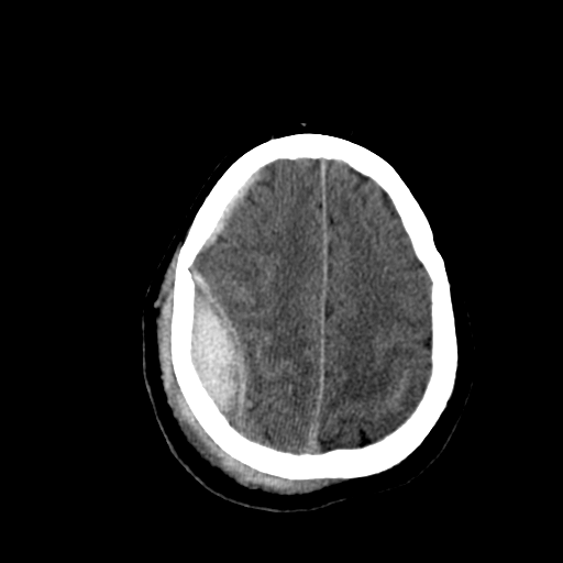
Epidural hematoma (EDH) – in between the dura and the arachnoid mater. They are fairly rare (0.5% of all TBI). The TC feature is a biconvex or lenticular hematoma, usually in the temporal or temporoparietal regions, and often results from a middle meningeal artery lesion. Classic clinical presentation is an awake patient until herniation occurs… “They talk and die!”. They all need to be drained fast!
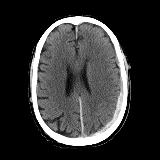
Subdural/subarachnoid hematoma (SDH) – in between the arachnoid and the pia mater. They are quite frequent. The TC feature is a semilunar shape that follows the brain’s contours. The issue here is not the hematoma itself, but the underlying parenchymal trauma. Surgical management comes if herniation of midline shifting is on the way.
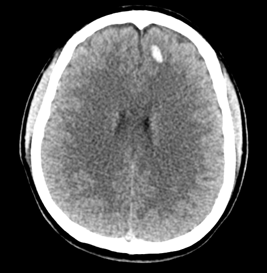
Cerebral contusions or Intraparenchymal Hemorrhage (IPH) – in the brain tissue itself, deeper than the pia mater. They are the commonest among the TBIs, the problem is they may have an evolution in hours or days: contusions can evolve to form an intracerebral hematoma or a coalescent contusion with enough mass effect to require immediate surgical evacuation. This is why almost all TBI repeat a CT scan 24-48 hours after the index trauma.
Now let’s go with what we have to do and pay attention to during our primary survey.
As we know, we evaluate the neurological state during 2 moments. We have to do it briefly in A (patient AVPU), and a bit more completely during D (GCS, pupils, and lateralizing signs).
If you don’t have it extremely clear, we suggest you read our primary survey’s posts.
Evaluating the GCS score is extremely important because it has a prognostic value for the patient. If you’re not very familiar with the GCS or you don’t practice it every day, here is a resource you should always have with you.
According to severity, we have to classify TBIs as:
- Mild (GCS 15-13)
- Moderate (GCS 12-9)
- Severe (GCS 8-3)
Always remember to evaluate the GCS of your patient frequently! It may change rapidly, and so will the aggressiveness of your treatment!
The Glasgow Coma Scale (GCS) and Its Implications
Let’s quickly review the management protocol for each grade:
Mild TBI (GCS of 15-13):
We are speaking about (semi)awake patients, so we have to complete our primary survey first and then move to our secondary one. During the AMPLE medical history, we should pay attention to signs of intoxication, amnesia, and confusion.
Remember to never ascribe an altered mental state to intoxication before completing the evaluation and the imaging!!!
But does every patient need a CT scan? Eventually not!
If the patient does not meet the Canadian CT head criteria (or the Canadian C-Spine criteria or the NEXUS ones), a CT scan may not be indicated.
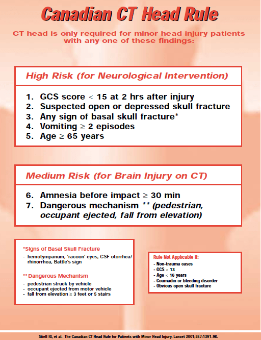
As you have to remember the CT scanning of the head and C-spine are connected, you cannot only scan one of the two. So, if the patient has no criteria for a CT head but has C-spine criteria, you’ll have to ask for both exams (the risk of connected injuries is extremely high)!
Indeed, if we do not have to scan the patient or if we have scanned it, after a few hours of observation, we may think about discharging the patient if NONE of the following items are checked:
- Abnormal CT scan
- Penetrating head injury
- History of prolonged loss of consciousness
- Deteriorating level of consciousness
- Moderate/severe headache
- Significant intoxication
- Skull fracture
- CSF leak (as rhinorrhea or otorrhea)
- Significant associated injuries
- No reliable companion at home
- GCS<15
- Neurological deficit
If none of the above are present, only 3% of those patients will evolve and be back to the hospital for a new evaluation. This is why not having a reliable companion during the 48 hours after the index trauma is an indication to hospitalize the patient.
Moderate TBI (GCS 12-9):
These are more or less the 15% of all TBIs we’ll see in A&E. Luckily, less than 20% of them will deteriorate to a coma. As always, we need to pass through the primary survey, proper resuscitation, secondary survey, CT scan, and neurosurgical consultation.
After that, almost all patients will repeat CT scans before discharge (if they get better) or immediately (if they deteriorate despite normal ABC).
Obviously, we have to think if we need to transfer the patient. If we have to, we should avoid the CT scan and not lose time!
Severe TBI (GCS 8-3):
Here we find the coma patients. As “A” we need to secure it, those patients need intubation in order to protect the airway in case of vomit, improve cerebral oxygenation, and control CO2. All those are treatments are fundamental to avoiding secondary brain injuries.
Clearly, as soon as those patients stabilize, they need a CT scan (remember that D comes after A, B, and C!).
Treatment
Finally, we can speak about treatment!
As we said, we need to prevent secondary brain injuries… This mainly means we have to restore normal perfusion, oxygenation, and function of the central nervous system. A “traumatized” brain loses its strength against adversity, and so, even a short hypotension or minor hypoxia may damage it!
Our aims are to keep/reach:
- Systolic BP >100 mmHg
- Temperature 36-38°C
- Glucose 80-180 mg/dL
- Hb >7 g/dL
- INR <1,4
- Na 135-145 mEq/l
- Sat O2 >95%
- PaO2 >99 mmHg
- PaCO2 35-45 mmHg
- ICP 5-15 mmHg
Basically, we need to restore normal physiology!
Now, before going into further treatment options you should have already called a neurosurgeon and shared the treatment plan!
We can do simple things in order to control a raised ICP (or a suspected one): loosen the cervical collar, raise the head of the patient at 30° or use a reverse-Trendelemburg position (if not contraindicated), and optimize pain control and sedation.
If the patient needs to be tubed (whatever the reason), ketamine is fine, it does not increase the ICP. Remember to sedate all your patients! Even a GCS of 3 may have a cough reflex that will make the ICP skyrocketing!
Mannitol can be used to decrease intracranial pressure thanks to its diuretic effect (1 g/kg) when the patient has a high ICP. Remember to not use mannitol in case of hypotension, it will only worsen it, in those cases, hypertonic saline will come in help (neurosurgeons or anesthesiologists will guide the use in this case). Unfortunately, there is still no strong evidence on this topic.
Barbiturates are effective in reducing ICP refractory to other measures, although they should not be used in the presence of hypotension or hypovolemia. So, they are not very suitable during the resuscitation phase.
Phenytoin or other anticonvulsants are no longer suggested as preventive treatments after TBI.
Lastly, we need to mention hypocapnia: it is done by hyperventilating the patient so the CO2 levels will drop between 25 and 30 mmHg. This will lead to intracranial vasoconstriction with decreasing ICP. Clearly, this will lead to hypoxia and ischemia in the brain tissue! We only need to use this treatment as a bridge while other therapies are on their way (mannitol, hypertonic saline, a scalpel, or a driller, etc…)
The brain cannot stand hypoCO2 for a long time!
The concept of “permissive hypotension” does not apply to TBIs patients, even in suspected ones! A normal physiologic blood pressure target should be targeted in this population.
Surgical decompression or evacuation (those are general indications, always consult a neurosurgeon for any doubt or patients with pathologic CT) are:
- Epidural hematomas >30 cm3 regardless of the GCS, or GCS <9 and anisocoria, or clot thickness >15 mm, or midline shift >5 mm, or neurologic deficit;
- Subdural hematomas >10 mm, or midline shift >5 mm, or GCS <9 or declining (≥2 points after admission), or anisocoria, or ICP >20 mmHg;
- Parenchymal lesions, progressive neurologic decline, mass effect, refractory ICP hypertension, GCS <9, frontal or temporal contusions >20 cm3, midline shift >5 mm, or any lesions >50 cm3.
In all severe TBIs, the ICP should be monitored (how to do it will depend on the neurosurgeon’s choice). The general goal is to maintain ICP <20 mmHg and CPP between 50-70 mmHg. Clearly, CPP and ICP are inexact surrogates of the cerebral blood flow meaning multimodal monitoring is always needed!
On top of all that, we will have to correct coagulopathy or correct anticoagulation treatments!
Brain Death
Brain death… We know, but we do have to cover this issue, or at least touch it…
Diagnosis of brain death requires meeting these criteria:
- Glasgow Coma Scale of 3;
- Nonreactive pupils;
- Absent brainstem reflexes (e.g. oculocephalic, corneal, and doll’s eyes, and no gag reflex);
- No spontaneous ventilatory effort on formal apnea testing;
- Absence of confounding factors such as alcohol or drug intoxication or hypothermia.
Many reversible conditions, like extreme hypothermia or barbiturate coma, can mimic brain death. Therefore, consider making this diagnosis only after all physiological parameters are normalized and central nervous system function is not potentially affected by other treatments.
As always, children are often able to recover from extremely severe brain injuries… So consider diagnosing brain death in these patients with extreme care!
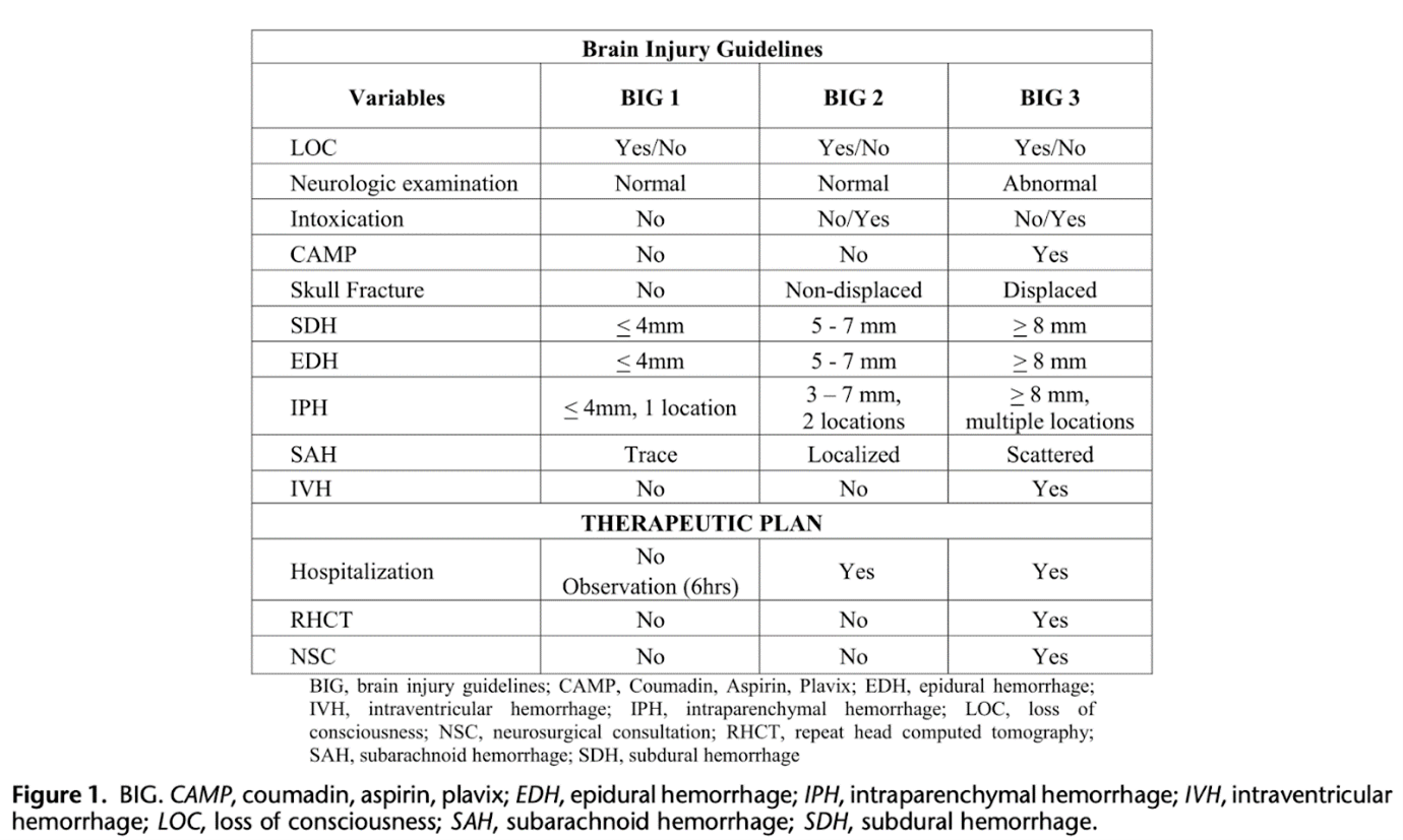
To close this post we have to cite the new BIG guidelines from 2014 recently validated by a multicenter study supported by Bellal Joseph and published in 2022.
After having a full evaluation and a CT scan, patients are divided into 3 groups according to severity and, consequently, to treatment strategy.
The main update from these “new” guidelines is the no necessity for neurosurgical consultation for patients in groups BIG 1 and 2, leaving the management to the acute care surgeon. The principle behind this is that only some of the patients in group 3 needed a neurosurgical intervention, and almost no patients in groups 1 and 2 required it.
How is this going to change things: up to now the first evaluation of TBI patients was pretty clear (i.e. ATLS protocols), but after the first CT scan we were still lacking good scientific-based protocols on how to manage patients during the first day/days after the index trauma. Maybe now we are finally heading to some widely validated pathways to manage our patients!
Now, let’s have a look at the table: basically, we would have to observe patients in BIG 1 for 6 hours and, if no changes in the neurological examinations are recorded, these patients could be safely discharged home. Similar management with BIG 2 patients, but with this group we need to admit them to prolong the observation for 24-48 hours. Again, if the neurological evaluation is normal after that timeframe, patients may be discharged home. If at any moment the neurological evaluation becomes altered, the patient jumps directly to BIG 3 and then a neurosurgical evaluation is needed!
Directly from the original article: “Patients categorized as BIG 3 had a severe head injury, and the optimal therapeutic plan for these patients consisted of hospitalization, an NSC, and a follow-up RHCT. Patients categorized as BIG 3 were on antiplatelet or anticoagulation medications, had an abnormal neurologic examination finding, and concerning CT scan findings (displaced skull fractures, and diffused ICH Q 8 mm). Nonexaminable patients, intubated patients, and patients with more than one CT scan finding were also categorized as BIG 3.”
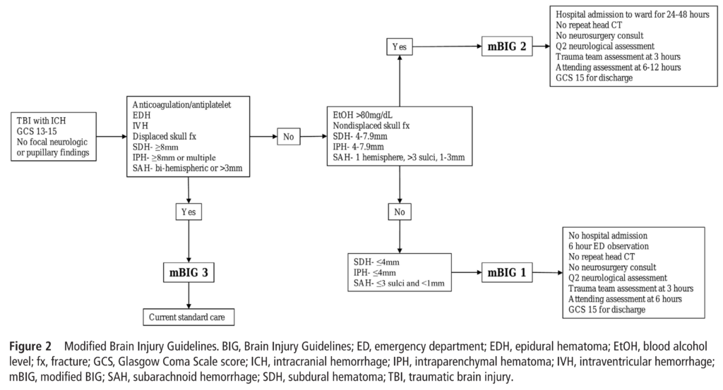
Another recent paper, published in 2020, has supported the use of the BIG guidelines for the initial management of TBI patients. However, the authors suggested some changes, mainly in the inclusion criteria in the BIG 3 group, considering all patients with an EDH as BIG 3 due to their high risk for clinical progression.
As always, we do really hope this was of some help to you!
Now we can get back to our patient… What would you do? We are speaking about a hemodynamic normal patient with a traumatic brain injury of what grade? What is your plan? In which BIG group do you think the patient would be?
We know you can solve this!
See you next time!
Namasté…
References
- Advanced Trauma Life Support: Student Course Manual. 10th Ed. Chicago, IL: American College of Surgeons; 2018.
- Vella MA, et al. Acute Management of Traumatic Brain Injury. Surg Clin North Am 2017;97:1015-30.
- Joseph B, et al. The BIG (brain injury guidelines) project: defining the management of traumatic brain injury by acute care surgeons. J Trauma Acute Care Surg 2014;76:965-9.
- Joseph B, et al. Validating the Brain Injury Guidelines: Results of an American Association for the Surgery of Trauma prospective multi-institutional trial. J Trauma Acute Care Surg 2022;93:157-65.
- Khan AD, et al. Multicenter assessment of the Brain Injury Guidelines and a proposal of guideline modifications. Trauma Surgery & Acute Care Open 2020;5:e000483.
How to Cite This Post
Marrano E, Bellio G. Newton & the Palm Tree: the Awful Coconut Law. Surgical Pizza. Published on November 26, 2023. Accessed on September 3, 2026. Available at [https://surgicalpizza.org/trauma/newton-and-the-palm-tree/].




3 Comments
Diego Visconti
Wow, such a nice and detailed chapter !
What about the last recommendations to associate a CT spine in all old patients (> 65 years old) who sustain a head injury (mild/moderate/severe, even with falls from their own height) due to the possibility of missed injuries in the C-spine ?
SurgicalPizza
Thank you!!! We will need to write at least an entire post on spinal injuries and indications for CT spine… Complex and interesting events…
According to the Canadian CT head rule, age >65 yo is a high-risk feature, warranting a CT head. In this case, the risk of synchronous associated injury to the C-spine is high, mandating a CT C-spine as well. BTW, age >65 yo is a high-risk feature even in the C-spine algorithm.
Pingback: